Digitization of X-rays inspection An easy to understand the scintillator
Digitization of X-rays inspection
In 1981, a method called Computed Radiography (CR) appeared, and has been driving the digitization of this inspection for about 20 years. However, CR had problems such as large equipment, long inspection time due to long image processing time, and large exposure dose.
In 1998 a Flat Panel Detector (FPD) device appeared, and immediacy and Detective Quantum Efficiency (DQE: the number of X-ray photons contributing to image formation, normalized by the number of incident X-ray photons per unit area) was improved. This leads to the inspection quality improvement and the acceleration of medical information digitization.

In the indirect conversion type FPD, which is one of the FPD methods, a light receiving element arranged in two dimensions (plane), and a scintillator are integrated. GOS (Gd2 O 2: Tb) and CsI (CsI: TI) are well known as scintillators used for X-rays inspection.

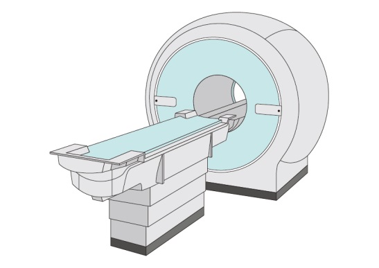
Furthermore, X-rays CT (Computed Tomography) came up by the discovery of the X-rays diffraction phenomenon in the 1910's, and the establishment of the radon transformation which is the mathematical foundation of tomography. X-rays tomography inspection has become possible, and diagnosis has become more sophisticated. The first X-rays CT equipment in the world was made by EMI Corporation in the UK in 1971. X-rays CT requires a fast data reading speed of the detector, so a scintillator with fast decay time of scintillation light is used.
With these advances, the digitization of X-rays examination has progressed, leading to digital information management at the medical field, and new examinations in combination with other examination methods are also being studied. X-rays CT has been applied not only to the medical field but also to industrial applications, and tomographic imaging in the foreign matter inspection and the defect inspection is also put to practical use.
- 1.What is scintillator?
- 2.Type of scintillator
- 3.Outline of X-rays inspection
- 4.Digitization of X-rays inspection
- 5.Radiation inspection other than X-rays examination
- 6.Keyword interpretation
